Imaging Services
Service Overview
Imaging is an important diagnostic tool that is used by many medical specialties.
Our imaging staff is expertly trained and certified to provide you with accurate, efficient imaging services in a comfortable, supportive environment.
General Conditions & Treatments
- Ultrafast/Electron Beam CT Scan
- Bone Scans
- Mammograms
- Nuclear Medicine
- Positron Emission Tomography (PET)
- Chest X-Ray
- Abdominal X-Rays
- X-rays of the Skull
- CT Screening Boosts Lung Cancer Survival
Patient Resources
What is angiography?
Angiography is an X-ray examination of the blood vessels that allows doctors to see how blood circulates within the body. It is used when a blockage in the flow of blood or abnormality of a blood vessel is suspected.
An angiogram can tell a doctor whether an artery is blocked, where the blockage is, how severe it is and what the cause is. A common cause of blockage is a blood clot in an artery narrowed by arteriosclerosis, or hardening of the arteries.
How does an angiogram work?
A radiologist will place a thin, flexible tube called a catheter into an artery or vein and then inject a small amount of contrast dye into the blood vessel to make it more visible in an X-ray.
What is the angiogram process?
There are just a few steps in this simple and nearly painless procedure:
- Patients are positioned on a comfortable table and X-rays are taken to get a pre-test picture.
- Patients are then given a local anesthetic where the catheter will be inserted. A small incision will be made and a thin catheter inserted into the artery.
- When the tube is exactly in place, some distance along the artery, the X-ray dye will be injected and the catheter removed. While the contrast dye is being injected, patients may experience a feeling of warmth, a metallic taste, or nausea. These symptoms are normal and should only last a minute or so.
- Removal of the catheter tube does not hurt, but pressure will be applied when it is removed to prevent the artery or vein from bleeding.
The advanced X-ray camera used in our angiography testing will show exactly where the dye reaches and where it doesn't; indicating if there is a blockage or abnormality in the blood vessel.
The entire process usually takes one to two hours to complete.
How do I prepare for my angiogram?
Patients receive a call to discuss what they can and cannot do before and after their test. However, as a general guide:
- Patients who are allergic to X-ray dye or iodine should let their doctor know as soon as possible.
- Smokers should not smoke for at least 24 hours before their angiogram as nicotine may affect the results of some tests.
- Patients who need to take medications in the morning should wash them down with just a few sips of water. They should also bring all medications with them to the hospital.
- Patients should not eat any solid food after midnight the night before their procedure. Patients may, however, have clear fluids, such as water or broth.
What happens after the test?
If a patient is having the procedure done as an outpatient, they will remain in the recovery room for four to six hours following your angiogram. Although they will be asked to lie still, they will be able to eat and drink. Visitors are allowed.
Someone will need to drive the patient home, and the patient should not drive for the rest of the day. Patients should also relax and take it easy at home. If they have a desk job, they should be able to return to work the next day but should avoid any lifting or straining for at least a week.
Before the patient goes home, the radiology nurse will give them further instructions.
When will I get the results?
A radiologist will read the scan and send a report to the physician within 24 hours. The patient’s doctor will then contact them to discuss their results.
Will my insurance cover angiography?
Most insurance plans cover this test. Patients should contact their insurance plan if they have any questions.
Advanced imaging technology can better detect diseases at an early stage, when a wider array of effective treatment options may be available.
Faster scanning time with the 320-slice CT scanner
- Technology is two to 10 times faster than most other CT scanners
- 320 images can be acquired rapidly, reducing what used to take minutes down to seconds
- Faster scanning time benefits patients, especially those with breathing difficulties or some other distress
- Speed and detail aid in decision-making in emergency cases
3D images provide enhanced diagnostic capability
Detailed information captured by the 320-slice CT scanner is used to generate 3D images with greater anatomical detail.
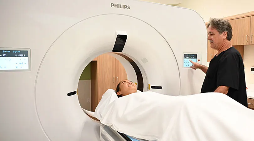
How is CT scan used?
CT scans provide detailed views of many types of tissue, including the lungs, bones, soft tissues and blood vessels and allow doctors to more effectively detect and treat a range of life-threatening illnesses, such as cancer and lung disease.
The unmatched speed and detail of the new 320-slice CT scanner also aids in decision-making in emergency cases where physicians have to make critical decisions quickly.
Diagnosis made with the assistance of CT can eliminate the need for invasive exploratory surgery and surgical biopsy and is effective in determining, at an early stage, the appropriate treatment for patients.
What happens during the scan?
During the non-invasive test, the patient is placed on a table and moved incrementally through the squared off donut-shaped scanner while an X-ray beam is projected through cross sections of their anatomy. The X-ray energy passes through the patient and is recorded on electronic detectors in the scanner. This information is then sent to a specialized computer that reconstructs the information into individual slices and combines them sequentially into a comprehensive volume image of the entire scanned area. The thinner the slices, the more revealing the detail is in the resulting images and the more definitive the exam results.
When will I get the results?
A radiologist will read a patient’s scan and send a report to their physician within 24 hours. Their doctor will contact them to discuss their results.
Will insurance cover a CT scan?
Most insurance plans cover this test. Patients should contact their insurance plan if they have any questions.
Will the scan hurt?
No, patients won't feel a thing. They simply lie still during the scan.
Is it safe?
MRI technology has been available for more than 20 years and is extremely safe. Unlike X-rays, MRI does not use radiation.
How long does it take?
Depending on each situation, an MRI could take between 30 and 60 minutes.
What happens during the scan?
It's very simple:
- Patients are positioned on the scanning bed.
- An instrument called an image coil will be placed over the area examined.
- The bed (tumbrel) will then be moved into the scanning chamber.
- Patients will be asked to lie very still during the scan.
- A doctor may request that a patient receive an injection of contrast agent to provide additional information about the area being scanned. The injection feels like any other shot, and is very safe.
- During the scan, patients may hear a chirping sound from the scanner. This is normal.
Can anyone have an MRI?
Since MRI is very safe, most people can have a scan done. However, if a patient has a pacemaker or metal implant, or if they are a woman in their first trimester of pregnancy, they cannot be scanned.
When will patients get the results?
A radiologist will read a patient’s scan and send a report to their physician within 24 hours. Their doctor will contact them to discuss their results.
Will insurance cover an MRI?
Most insurance plans cover MRI. Patients should contact their insurance plan if they have any questions.
How should a patient prepare for their scan?
- Patients should arrive 30 minutes before their scheduled appointment and go to the Registration Department.
- Patients should dress in warm, comfortable clothing (sweatshirt and pants work well).
- Patients cannot wear anything metallic during the exam, so it's best for them to leave watches, jewelry or anything else containing metal at home.
- If sedation is ordered, a doctor will provide further instructions.
Care at Kapiʻolani
Kapiʻolani Medical Center for Women & Children is Hawaiʻi’s leader in pediatric and women’s health. Our Imaging Department specializes in diagnostic services for newborns, children and teens, while also providing general imaging for select adult studies. We combine state-of-the-art technology with a compassionate, family centered approach to ensure accurate results in a safe, supportive environment.
Our department provides a full range of imaging modalities:
- X‑Ray (Radiography): Quick, low‑dose exams that capture detailed images of bones, chest, and other body structures. We are uniquely equipped to provide specialty pediatric imaging.
- Fluoroscopy: Real‑time imaging that allows physicians to observe movement inside the body, such as swallowing studies or gastrointestinal exams.
- CT (Computed Tomography): Advanced cross sectional imaging. We use pediatric‑specific protocols to minimize radiation exposure while ensuring clear results.
- Ultrasound: Safe, radiation free imaging for soft tissues and organs.
- Echocardiography (Echo): Specialized ultrasound of the heart.
- MRI (Magnetic Resonance Imaging): Detailed imaging using magnetic fields, with child friendly protocols and sedation options when needed. Our staff are highly specialized in pediatric and breast MRI.
- Nuclear Medicine: Functional imaging using small amounts of radiotracers to evaluate organ function and detect disease.
We also provide Interventional Radiology services for both children and adults. Treatments offered include:
Vascular Malformations
- Sclerotherapy of venous malformations, lymphatic malformations and arteriovenous malformations
- Multidisciplinary treatment with the Vascular Anomalies Clinic
Gynecologic Interventions
- Uterine artery embolization for treatment of abnormal uterine bleeding (e.g. fibroids and adenomyosis)
Enteral Access
- Percutaneous gastrojejunostomy (GJ) tube placement and maintenance
- Percutaneous gastrostomy (G) tube placement
Vascular Access
- Implanted venous ports (Mediports)
- Peripherally inserted central catheters (PICC)
- Tunneled central venous catheters including hemodialysis catheters
- Temporary central venous catheters
Biopsies
- Image guided biopsies
- Fine needle aspiration
Drainages
- Abscess drainage
- Chest tubes, fibrinolytic therapy and thoracentesis
Other
- Arthrograms and joint injections
- Bile duct evaluation, biliary drains and gall bladder drains
- Varicocele embolization
- Lumbar punctures
- Lymphangiography and lymphatic system interventions
- Nephrostomy tube, ureteral stents and cystostomy tube placement
- TMJ injections
Care at Pali Momi
Pali Momi Medical Center is a leader in innovation and patient-centered care. As the first in West Oʻahu to introduce a permanent MRI, a 320-slice CT scanner, and a cardiac catheterization program, we continue to set the standard for advanced diagnostic and interventional services. We offer state-of-the-art technology for accurate, timely diagnosis, maintain radiation doses well below national standards for patient safety, and provide convenient scheduling—including early mornings and weekends—all within a comfortable, patient-focused environment.
Comprehensive Imaging Services
- CT Imaging – We provide a full range of CT services, including CT Lung Screening, Cardiac CT, and stroke imaging as part of our Stroke Care Center designation. Our team also performs CT-guided biopsies along with general CT studies, ensuring precision and timely diagnosis for complex conditions.
- Diagnostic Radiology - Our highly skilled technologists, trained to Trauma II standards, work closely with our expert radiologists to deliver precise, high-quality imaging across a wide range of conditions. This collaboration supports specialized care, including our Bone and Joint Center, ensuring accurate diagnosis and treatment planning. We also provide fluoroscopy for dynamic imaging studies, offering real-time visualization to guide complex procedures safely and effectively.
- MRI Services – Including 1.5T and 3T MRI systems, we deliver superior image clarity for complex exams such as comprehensive breast MRI and high-resolution prostate MRI studies—ensuring accurate diagnosis and treatment planning. To enhance patient safety, we have an MRI Safety Officer on staff who coordinates care for patients with metal implants, providing expert guidance and peace of mind.
- Nuclear Medicine Diagnostics – Our team of nuclear medicine technologists and radiologists carefully evaluates each case to determine the most appropriate diagnostic study or specialized treatment. This collaborative approach ensures accuracy and personalized care. Services include Cardiac Stress Tests, Myocardial Perfusion Imaging, Gastric Emptying Studies, HIDA Scans, Bone Scans, Lung Scans, GI Bleed Studies, Renal Scans, Lymphoscintigraphy, and Thyroid Uptake Exams—all designed to provide precise answers for complex conditions.
• Ultrasound – Our skilled team—holding multiple advanced certifications—provides high-quality imaging for general, abdominal, renal, thyroid, and vascular studies. We also perform ultrasound-guided biopsies in collaboration with radiologists and offer UroNav prostate biopsy, delivering targeted precision for prostate care.
• Interventional Radiology (IR) - Our dedicated IR Suite offers minimally invasive, image-guided procedures for vascular, oncologic, and other targeted therapies—designed to reduce recovery time and improve outcomes. Additionally, our IR services include prostate and uterine embolization’s, nephrostomy tube placement, gastrostomy, biliary drainage, and biliary rendezvous procedures.
• Pali Momi Women’s Center - Conveniently located near Pearlridge Center, the Pali Momi Women’s Center offers a spa-like environment dedicated to women’s health. Services include 3D mammography, breast ultrasound and non-surgical biopsy, and a comprehensive Bone Health Program with DEXA scanning. Patients also benefit from dedicated navigators and cancer support programs, ensuring compassionate care every step of the way.
• Radiologists - Our team of radiology physicians combines fellowship-trained interventional specialists with subspecialists dedicated to precision and innovation. From minimally invasive procedures to advanced neurological imaging, we provide care that improves outcomes. With expertise in breast and prostate imaging, orthopedic diagnostics, and nuclear medicine, we use state-of-the-art technology to support early detection and personalized treatment. Our radiologists bring decades of experience to every image, delivering clarity and confidence in every diagnosis. We don’t just interpret—we uphold the highest standards in medical imaging.
Experience advanced imaging and women’s health care with compassion and expertise.
Pali Momi Imaging Services: 808-535-7733
Care at Straub Benioff
At Straub Benioff Medical Center, we provide a full spectrum of imaging services from routine X-rays to the most advanced interventional radiology procedures all designed to deliver answers with precision and care.
- General X-rays: Produces high quality X-rays to support all providers especially the Bone and Joint Center.
- MRI Excellence: Our new 1.5T MRI system coupled with our highly skilled staff safely accommodates nearly every type of exam that isn’t contraindicated, offering clear, detailed images to guide diagnosis and treatment.
- Comprehensive CT Imaging: Our CT technologists specialize in complex exams across cardiology, orthopedics, ENT, neurology, gastrointestinal, and genitourinary specialties.
- General Ultrasound Expertise: Safe and radiation-free, ultrasound helps providers evaluate organs such as the liver, kidneys, gallbladder, and thyroid with ease and accuracy.
- Complex Vascular Lab Imaging: Using painless ultrasound, our technologists assess blood flow in veins and arteries to detect blockages or narrowing all without radiation or needles.
- Women’s Services: We offer digital mammography, tomosynthesis, breast ultrasound, MRI breast exams, and DEXA (bone density) scans. Straub Benioff proudly features the only state’s upright stereotactic breast biopsy system, allowing patients to undergo biopsies in a comfortable seated position.
- Nuclear Medicine Innovation: Our Nuclear Medicine Department provides advanced radioligand therapies such as Pluvicto, Lutathera, and Xofigo, alongside diagnostic studies including HIDA scans, gastric emptying exams, and cardiac stress tests.
- Interventional Radiology: Our team performs the widest range of vascular and non-vascular procedures within the system including but not limited to cerebral angiography, aneurysm coiling, embolization, nephrostomy tube placement, gastrostomy, biliary drainage, biliary rendezvous, and TIPS procedures.
- Expert Radiologists: More than 10 fellowship-trained radiologists bring subspecialty expertise in neuroradiology, musculoskeletal imaging, cardiac imaging, breast imaging, interventional radiology, and body imaging.
Straub Benioff combines cutting-edge technology with compassionate care ensuring every patient receives the answers they need, delivered by experts they can trust.
Straub Benioff Imaging Services: 808-522-4221
Care at Wilcox
The center is equipped with state-of-the-art technology to serve patients across the inpatient, outpatient, and emergency settings.
The spectral-detector CT scanner produces comprehensive images for patients of all ages, including children, and with a range of medical needs, such as bariatrics. The machine generates high-quality, exceptionally clear images with faster setup times and shorter exam times. This results in a lower dose of radiation as well as a more comfortable overall experience.
The 320-slice CT scanner offers advanced imagery full-body scanning in a single rotation of the X-ray tube. This faster scan time is helpful for patients who have a hard time staying still for extended periods.
Advanced features of the CT scanners at Wilcox help medical teams detect, diagnose and monitor both common and chronic diseases, disorders and conditions, including cancer, heart disease and bone fractures.
Imaging services are available 24 hours a day, seven days a week. Other services offered include X-ray; Ultrasound; Mammography; MRI; Nuclear Medicine and limited interventional radiologic procedures. Board-certified physician radiologists provide quality care and timely interpretation of radiographic procedures.
Services are by referral from a patient’s primary care physician, who will use these internal images of the body to assist with diagnosis and treatment.
Wilcox Family Imaging Center: 808-245-1030
Our Imaging Services Locations
Oʻahu
Pali Momi Medical Center Pali Momi Medical Center 98-1079 Moanalua Road ‘Aiea, HI 96701 Phone: 808-486-6000 Get directions Details Pali Momi Outpatient Center Pali Momi Outpatient Center
98-1005 Moanalua Rd.
Aiea, HI 96701
Phone: 808-486-6000
Get directions
Details
Pali Momi Women's Center
Pali Momi Outpatient Center
98-1005 Moanalua Rd.
Aiea, HI 96701
Phone: 808-486-6000
Get directions
Details
Pali Momi Women's Center
 Pali Momi Women's Center
98-1005 Moanalua Road
Aiea, HI 96701
Phone: 808-485-4500
Get directions
Details
Straub Benioff Medical Center – Kāhala Clinic & Urgent Care
Pali Momi Women's Center
98-1005 Moanalua Road
Aiea, HI 96701
Phone: 808-485-4500
Get directions
Details
Straub Benioff Medical Center – Kāhala Clinic & Urgent Care
 Straub Benioff Medical Center – Kāhala Clinic & Urgent Care
4210 Waiʻalae Ave., Suite 501
Honolulu, HI 96816
Phone: 808-462-5300
Get directions
Details
Straub Benioff Medical Center – Kāhala Clinic & Urgent Care
4210 Waiʻalae Ave., Suite 501
Honolulu, HI 96816
Phone: 808-462-5300
Get directions
Details
Kauaʻi
Wilcox Medical Center Wilcox Medical Center
3-3420 Kūhiō Highway
Līhu‘e, HI 96766
Phone: 808-245-1100
Get directions
Details
Wilcox Medical Center
3-3420 Kūhiō Highway
Līhu‘e, HI 96766
Phone: 808-245-1100
Get directions
Details